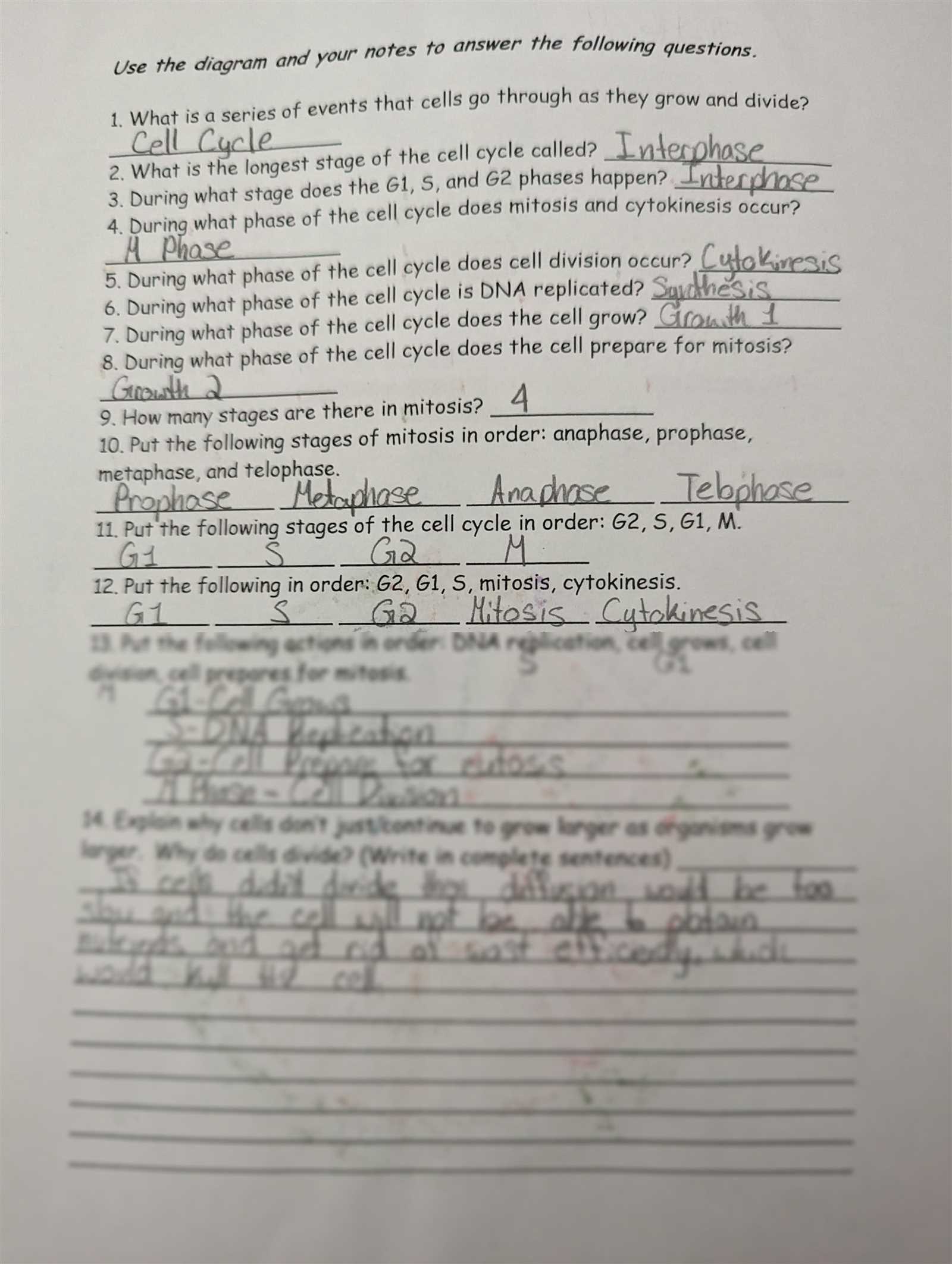
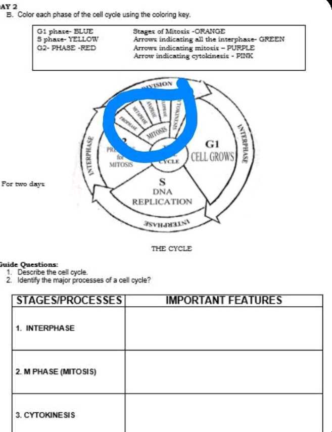
Mitosis Coloring Homework Answers and Tips

Understanding the process of cell division is a crucial part of biology. This topic explores how cells reproduce and create new ones, ensuring growth and tissue repair. The stages involved in this process are fundamental to the study of life, and grasping their details helps to build a stronger foundation in cell biology.
When tackling assignments related to this subject, it’s important to approach the task systematically. Clear visualization of each phase is key to comprehending the overall mechanism. By following a structured method, students can improve their understanding and avoid common pitfalls that can arise from confusion or incomplete knowledge.
In this guide, we will provide essential insights into how to break down these stages and present them accurately. With practical advice and helpful tips, you’ll be able to tackle this topic with confidence and precision. Mastering the key concepts will not only enhance your academic performance but also deepen your appreciation of cellular processes.
Cell Division Visualization Assistance
When studying cell division, illustrating each stage accurately plays a vital role in understanding the process. Properly mapping out the different phases allows students to see the transitions and transformations that occur within a single cell. By representing these stages visually, the complexities of the division process become easier to grasp.
Key Phases to Focus On
- Interphase – The cell prepares for division by growing and duplicating its DNA.
- Prophase – Chromosomes condense, and the nuclear membrane begins to break down.
- Metaphase – Chromosomes align at the cell’s equator, preparing for separation.
- Anaphase – Chromatids are pulled apart to opposite sides of the cell.
- Telophase – Two new nuclear membranes form around the separated chromatids.
- Cytokinesis – The cell divides into two distinct daughter cells.
Effective Techniques for Representation
- Choose contrasting colors to distinguish between chromosomes, centromeres, and other key components.
- Use clear labels for each stage to guide your visualization and ensure accuracy.
- Take your time to carefully depict the changes that occur in each phase for clarity.
By following these methods, you will develop a clear and thorough representation of the division process. This technique not only enhances your comprehension but also improves your ability to recall key details when discussing or testing on the subject.
Understanding the Stages of Cell Division
Cell division is a highly organized and regulated process that ensures the proper replication and distribution of genetic material. The sequence of events involves several distinct phases, each with specific roles and characteristics. A clear understanding of these stages is essential for grasping how organisms grow, repair, and maintain their cells.
Overview of Key Phases
- Interphase – This preparatory phase includes growth and DNA replication, setting the stage for the cell’s division.
- Prophase – The chromatin condenses into visible chromosomes, and the nuclear membrane begins to dissolve.
- Metaphase – Chromosomes align along the center of the cell, preparing to be split into two new sets.
- Anaphase – The chromosomes are separated and pulled toward opposite poles of the cell.
- Telophase – New nuclear membranes form around the separated sets of chromosomes, signaling the near end of division.
- Cytokinesis – The final separation of the cell into two daughter cells occurs, completing the division process.
Significance of Each Stage
Each stage of division plays a critical role in ensuring that the daughter cells receive the correct amount of genetic material. Disruptions during any phase can lead to errors in cell function, which can have serious implications for the organism. A solid understanding of each stage helps to reinforce the importance of precise cellular processes in maintaining life.
How to Color Cell Division Effectively
Accurately illustrating the process of cell division requires more than just applying colors. The goal is to enhance understanding by highlighting key components and stages in a clear, organized manner. By choosing the right colors and techniques, you can make each phase stand out, helping to reinforce the concepts behind them.
Choosing the Right Colors
- Use contrasting colors for different structures, such as chromosomes, centrioles, and the nuclear membrane, to make each element easily identifiable.
- Pick bright colors for phases like prophase and metaphase, where key activities such as chromosome alignment are most visible.
- Choose more subdued shades for stages like telophase and cytokinesis to reflect the calming transition toward cell separation.
Organizing Your Approach

- Start by carefully outlining each part of the diagram before applying color, ensuring all structures are visible and correctly placed.
- Label each phase clearly to avoid confusion and ensure you are depicting the right stage of the division process.
- Work systematically from one stage to the next, using consistent color schemes throughout for clarity.
By following these steps, you’ll not only create an effective visual representation of the division process but also deepen your understanding of each phase. This method helps make the complex process easier to study and recall when needed.
Key Features to Highlight in Cell Division
When illustrating the process of cell division, it’s crucial to emphasize specific features that define each phase. Focusing on these key components allows for a clearer understanding of how cells replicate and distribute genetic material. By recognizing and accentuating these elements, you can create a more accurate and informative representation of the process.
Important Structures to Emphasize
- Chromosomes – These are the primary structures that carry genetic information. Their visibility and alignment are central to understanding the process.
- Centrioles – These structures help organize the microtubules during cell division, playing a key role in the separation of chromosomes.
- Nuclear Membrane – The breakdown and reformation of the nuclear membrane marks important transitions in the division process.
- Spindle Fibers – These are the threads that help pull chromosomes apart, ensuring the correct distribution of genetic material.
Phases to Focus On
- Prophase – The beginning of visible chromosome formation and the disintegration of the nuclear membrane.
- Metaphase – Chromosomes line up at the cell’s equator, preparing for division.
- Anaphase – Chromatids are pulled apart, moving toward opposite poles.
- Telophase – New nuclear membranes form, marking the near completion of division.
By paying attention to these critical features, you’ll be able to create a more detailed and accurate depiction of the cell division process. This will help reinforce your understanding of the mechanisms involved in cellular reproduction.
Common Mistakes in Cell Division Assignments
When studying the process of cell division, students often make mistakes that can lead to confusion and misunderstandings of the material. These errors typically arise from overlooking key details, misrepresenting the sequence of events, or failing to accurately depict the structures involved. Recognizing and avoiding these common mistakes is essential for mastering the subject and achieving a better understanding of how cells replicate.
Typical Errors to Avoid
- Incorrect Sequence of Phases – Failing to arrange the stages in the correct order is a frequent mistake. It’s crucial to understand the chronological flow from one phase to the next.
- Mislabeling Structures – Confusing key components, such as chromosomes and centromeres, or neglecting to label them properly can lead to inaccuracies in your representation.
- Overlooking Small Details – Skipping minor but important features, like the spindle fibers or nuclear membrane breakdown, can make the diagram incomplete.
- Inaccurate Color Usage – Using the wrong colors for different structures or phases can cause confusion and make it difficult to identify each part of the division process.
How to Avoid These Mistakes
- Study the process in depth to understand the sequence of events and the role of each structure.
- Carefully label each part of the diagram and double-check for accuracy.
- Focus on the smaller details, such as the movement of chromosomes or the formation of the nuclear envelope.
- Use a consistent and logical color scheme to clearly differentiate between different phases and structures.
By paying attention to these common pitfalls and taking the time to review your work, you can improve the accuracy of your cell division assignments and avoid confusion. A well-organized and detailed depiction will make it easier to understand the complex process of cellular reproduction.
Helpful Resources for Cell Division Visualization
When working on assignments related to cell division, using the right resources can significantly enhance understanding and accuracy. Various tools and materials are available that provide visual aids, detailed explanations, and interactive features to help visualize each stage of the process. Leveraging these resources can make the task more manageable and improve comprehension of this complex biological concept.
Recommended Resources
- Interactive Diagrams – Websites and apps that offer interactive cell division diagrams allow you to click through each phase and understand the transitions more clearly.
- Video Tutorials – Educational videos can walk you through the stages of cell division, highlighting key features and showing real-life examples.
- Textbooks and Study Guides – Well-organized study materials often include detailed illustrations of the division process with step-by-step breakdowns of each stage.
- Online Quizzes – Interactive quizzes can test your knowledge on the subject, reinforcing learning and helping to identify areas for improvement.
Where to Find These Resources
- Check educational platforms like Khan Academy or Coursera for free courses and tutorials.
- Explore websites dedicated to biology education, such as BioMan Biology or Learn Genetics, for interactive activities and resources.
- Visit your school’s online library or a trusted academic site for textbooks and study guides with detailed illustrations.
- Use YouTube to find educational channels like CrashCourse and Amoeba Sisters, which offer engaging videos on cell biology.
By utilizing these resources, you can gain a deeper understanding of cell division, enhance your visual representations, and ensure you have a comprehensive grasp of each phase. These tools will not only make the learning process easier but also more engaging and effective.
Importance of Cell Division in Growth and Reproduction
Cell division is a fundamental biological process that allows organisms to grow, repair damaged tissues, and reproduce. It ensures that new cells are produced with the same genetic material as the original, maintaining the integrity of the organism’s genetic code. This process is vital for the continuity of life and supports various functions essential for survival, such as growth, development, and healing.
Role of Cell Division in Organisms
Cell division plays a critical role in many aspects of life. It supports the growth of multicellular organisms, helps in tissue regeneration, and allows for the formation of gametes in reproduction. Without proper division, cells would not be able to replicate and pass on genetic information accurately, leading to problems like genetic mutations and tissue dysfunction.
| Function | Explanation |
|---|---|
| Growth | Cell division is crucial for the growth of an organism, allowing it to develop from a single cell into a complex system of tissues and organs. |
| Repair | When cells are damaged, division helps to produce new, healthy cells that replace the injured ones, aiding in tissue repair. |
| Reproduction | In organisms that reproduce sexually, cell division is key to the production of gametes, ensuring the passing of genetic material to the next generation. |
| Genetic Stability | Accurate division ensures that genetic material is copied and distributed evenly between new cells, preventing genetic errors. |
Why Accurate Cell Division is Crucial
Accurate and regulated cell division is necessary to avoid genetic anomalies that could affect the health of the organism. Errors during division can result in diseases such as cancer, where cells divide uncontrollably. Thus, understanding and maintaining the proper mechanisms of division is essential for the well-being of all living organisms.
Step-by-Step Guide for Cell Division Visualization
Creating a detailed and accurate representation of cell division involves a structured approach. By following a step-by-step guide, you can clearly distinguish between the different stages and structures involved in the process. This approach ensures that each phase is represented with precision, helping to reinforce your understanding of how cells divide and replicate.
Preparation Steps
- Gather Materials – Make sure you have all necessary materials, such as colored pencils, markers, and a diagram of the division process.
- Familiarize Yourself with the Stages – Review the key stages of division to understand what each phase looks like and the critical components involved.
- Choose a Color Scheme – Select colors that will clearly differentiate the various structures (e.g., use different colors for chromosomes, spindle fibers, and the nuclear membrane).
Step-by-Step Instructions
- Start with Prophase – Begin by coloring the chromosomes, which are usually condensed and visible. Use a bold color like red or blue for easy identification.
- Move to Metaphase – Color the chromosomes aligned at the cell’s equator. Highlight the spindle fibers that help pull the chromosomes apart.
- Proceed to Anaphase – Color the separating chromatids, ensuring they are distinct from one another. This stage marks the movement of chromosomes toward opposite poles.
- End with Telophase – Finally, color the new nuclear membranes forming around the separated genetic material, indicating the near completion of the process.
By following these steps, you will create a clear and organized representation of cell division, which will not only aid in visualizing the process but also help solidify your understanding of each stage involved in cellular replication.
Color Coding the Phases of Cell Division
Using color coding to differentiate the various stages of cell division is an effective strategy for both learning and visualizing this complex biological process. Each phase has distinct characteristics, and assigning specific colors to these phases helps to highlight their unique features, making the division process easier to follow and remember.
By using different colors for each stage, you can better understand how the cell progresses through its cycle. This method also makes it easier to distinguish between critical components like chromosomes, the cell membrane, and the spindle fibers, which play essential roles in the division process. Color coding provides a clear, visual representation that can enhance comprehension and retention of the material.
For instance, you might choose a color like blue to represent chromosomes, red for the spindle fibers, and green for the nuclear envelope. This approach not only improves the clarity of your visual diagram but also helps you quickly identify which structures belong to which phase. The use of color coding also supports active learning by encouraging engagement with the material in a more dynamic and interactive way.
Tips for a Successful Cell Division Assignment
Completing an assignment related to the process of cell division requires a focused approach and attention to detail. By following a few essential tips, you can ensure that your work is accurate, organized, and demonstrates a clear understanding of the stages involved. These strategies will help you create a comprehensive and effective representation of the process.
Understand the Key Stages
Before starting your task, take the time to study the stages of cell division thoroughly. Knowing the unique characteristics and events that occur in each phase will allow you to highlight and represent them correctly. Familiarizing yourself with the sequence of events also ensures that you can logically organize your work and make meaningful distinctions between each step of the process.
Organize Your Materials and Workspace
A tidy and well-organized workspace can make the task easier and more enjoyable. Gather all necessary tools, such as colored pencils, markers, and reference diagrams, before you begin. Keeping everything in its place will help you stay focused and avoid unnecessary distractions. Additionally, using a clean, clear diagram as a reference ensures that you can work efficiently and correctly.
Lastly, don’t forget to double-check your work. Review each phase for accuracy, ensuring that the stages are represented in their proper sequence and that each structure is colored according to your chosen scheme. This careful attention to detail will help you produce high-quality work that reflects your understanding of the material.
Understanding Chromosomes in Cell Division
Chromosomes are essential structures that carry genetic information within a cell. During the process of cell division, these structures undergo significant changes that allow the cell to replicate its genetic material and distribute it evenly to the daughter cells. Understanding the role of chromosomes is crucial for grasping how the process of cellular reproduction ensures genetic continuity and stability across generations.
In the early stages of division, chromosomes condense and become visible under a microscope, making it easier to track their movement. As the process progresses, chromosomes align, separate, and eventually reach the poles of the cell, ensuring that each daughter cell receives an identical set of genetic material. The precise behavior and organization of chromosomes during this process are vital to maintaining proper cellular function and preventing genetic errors.
Each chromosome is made up of DNA and proteins, forming a structure known as chromatin. During division, this chromatin condenses into tightly packed chromosomes, which are then divided between the two new cells. This ensures that both daughter cells receive a complete and accurate copy of the genetic blueprint needed for their growth and function.
Tools You Need for Cell Division Representation
To effectively visualize and represent the stages of cell division, having the right tools is essential. Whether you’re creating a diagram, a model, or completing an assignment, the materials you choose will significantly influence the quality and clarity of your work. Below are some of the essential tools that will help you accurately depict the key processes involved in cell division.
Basic Supplies
- Colored Pencils or Markers: Use these to differentiate between various stages and structures, making each part of the process easier to identify.
- Reference Diagrams: A clear, labeled diagram serves as a guide for understanding and accurately representing each phase.
- Paper or Poster Board: A sturdy surface to work on allows for better control and neatness, especially when dealing with larger projects.
- Pens or Fine-Tipped Markers: These are useful for outlining important structures like chromosomes, spindle fibers, and the cell membrane.
Advanced Tools (Optional)
- Digital Tools: If you’re creating a digital representation, software like drawing programs or specialized biology apps can help you produce high-quality visuals.
- Templates: Pre-drawn templates or outlines can help guide your drawing and ensure accuracy when representing complex stages.
By using the right combination of these tools, you can enhance your ability to depict cell division clearly and accurately. These resources will not only improve the presentation of your work but also deepen your understanding of the biological processes involved.
Best Practices for Cell Division Diagrams
Creating clear and accurate diagrams of cellular processes is essential for understanding complex biological concepts. When illustrating stages of cell division, following best practices ensures that the representation is both educational and easy to comprehend. The right approach not only helps in depicting the key structures but also aids in organizing information in a way that enhances learning.
Use Clear Labels and Annotations
Each component of the diagram should be clearly labeled to ensure that anyone viewing the diagram can easily identify different structures. Mark key elements such as chromosomes, spindle fibers, the nuclear envelope, and the cell membrane. Using legible, neat text and ensuring that labels do not overlap will make your diagram more readable. Annotations explaining each phase can further clarify the function of each structure during the process.
Maintain Accurate Proportions
While it’s tempting to exaggerate certain features for clarity, maintaining accurate proportions is essential for creating a scientifically sound diagram. Ensure that the size of the cell, chromosomes, and other structures are proportionate to one another. This not only improves the overall aesthetic but also reflects the true scale and relationships between different components, providing a more realistic and informative representation.
By following these best practices, you can create diagrams that are not only visually appealing but also educational, helping to simplify complex biological processes and enhance understanding.
How to Approach Cell Division Assignments
When tasked with illustrating or explaining the process of cell division, having a systematic approach is key to ensuring clarity and accuracy. Breaking down the assignment into manageable steps can help you focus on key concepts, organize your thoughts, and produce a comprehensive and well-structured result. Below are some effective strategies for tackling assignments on cellular division.
Step-by-Step Process
- Understand the Concept: Start by reviewing the key phases involved, such as the stages of division, the structures involved, and the overall sequence of events. Make sure you grasp the biological significance behind each phase.
- Gather Necessary Tools: Collect your materials, including colored pencils, diagrams, and reference books or online resources. Ensure you have everything required before you begin working.
- Create an Outline: Before you start drawing or writing, outline the main points that need to be covered. This will help you organize your assignment and ensure you include all necessary information.
Key Areas to Focus On
When working on your project, make sure to pay attention to the following important areas:
| Key Area | What to Focus On |
|---|---|
| Cell Structures | Accurately represent structures like chromosomes, spindle fibers, and the nuclear membrane. |
| Sequence of Events | Ensure that the stages are arranged in the correct order, from the initial phase to the final division. |
| Clarity and Detail | Provide clear labels and explanations to make the diagram or explanation easily understandable. |
By approaching the assignment with a structured plan and focusing on the most important aspects, you’ll be able to complete the task efficiently and accurately, ensuring that your work is both informative and well-presented.
Identifying Key Phases in Cell Division
Understanding the stages of cellular reproduction is essential for accurately depicting the process. Each phase of cell division plays a unique role in ensuring that genetic material is distributed correctly to two daughter cells. In this section, we’ll explore the key stages of cell division and highlight the important features to recognize during each phase.
Major Phases of Division
- Interphase: This phase precedes the actual division process, where the cell prepares for division by growing and duplicating its DNA. It consists of three sub-stages: G1, S, and G2.
- Prophase: The first stage of division, where the chromatin condenses into visible chromosomes, and the nuclear envelope begins to break down.
- Metaphase: The chromosomes align at the cell’s equatorial plane, preparing for separation. This phase ensures that the chromosomes are evenly distributed.
- Anaphase: The sister chromatids are pulled apart by spindle fibers towards opposite poles of the cell.
- Telophase: The chromatids reach the poles, and the nuclear envelope re-forms around each set of chromosomes, marking the end of the division process.
- Cytokinesis: The final step where the cell’s cytoplasm divides, forming two distinct daughter cells. This phase overlaps with telophase in many cells.
Recognizing Key Features in Each Stage
- Chromosome Behavior: During prophase, metaphase, and anaphase, you should be able to clearly identify how chromosomes behave and move within the cell.
- Spindle Formation: Look for the appearance of spindle fibers in prophase and their role in pulling the chromosomes apart during anaphase.
- Nuclear Changes: Pay attention to how the nuclear envelope disassembles and reforms during different stages, signaling the start and end of cell division.
By recognizing these distinct features in each phase, you will be able to better understand the process and accurately depict the events of cell division, ensuring a clear and detailed representation of the entire cycle.
How to Avoid Common Coloring Errors
When illustrating or labeling stages of cellular processes, it’s easy to make mistakes if the details are overlooked. The key to avoiding errors lies in understanding the process and applying careful attention to the specific features of each phase. Below are some strategies for ensuring accuracy while working on these types of diagrams.
Common Mistakes to Watch For
| Common Mistake | How to Avoid It |
|---|---|
| Misidentifying Chromosome Position | Make sure to pay close attention to the alignment of chromosomes, especially during metaphase. They should line up at the center, not be scattered. |
| Skipping Over Interphase | Ensure you represent interphase properly, as it is a preparatory phase. It’s easy to overlook, but it’s crucial for understanding cell division. |
| Incorrect Spindle Fiber Representation | Spindle fibers appear during prophase and help separate chromosomes in anaphase. Clearly show their connection to chromosomes to avoid confusion. |
| Overlooking the Nuclear Envelope | The nuclear envelope dissolves in prophase and reforms in telophase. Keep track of its changes, and avoid missing these important transitions. |
Tips for Precision
- Use Clear Color Codes: Designate specific colors for each phase, such as blue for interphase, red for prophase, and so on. Consistency is key to clarity.
- Check Chromatid Movement: Ensure that chromatids are separated properly during anaphase, and that they are still connected during metaphase.
- Double-Check Alignment: Before finalizing the diagram, review the arrangement of chromosomes and other structures to make sure they reflect the correct positioning in each stage.
By following these steps and paying close attention to details, you can avoid common errors and produce accurate, clear illustrations of the process.
Final Review of Mitosis Homework

Once you’ve completed your diagram or activity focusing on cell division, it’s crucial to conduct a thorough review to ensure accuracy and clarity. This step helps to verify that all phases are correctly identified and that no important details are missed. The final check is essential for reinforcing your understanding and ensuring your work reflects the correct biological processes.
Checklist for Final Review
- Verify Phase Order: Make sure the sequence of stages follows the correct progression, from the initial phase to the final one. Each phase should transition smoothly into the next.
- Double-Check Chromosome Placement: Ensure chromosomes are correctly placed within the appropriate stage. They should be well-defined and positioned according to the current phase.
- Examine Cell Structures: Look over structures like the nuclear membrane, spindle fibers, and centromeres. Verify that these features are appropriately represented for each phase.
- Review Color Coding: If using colors to distinguish different stages, check that the color scheme is consistent and each phase is easily identifiable.
Common Oversights to Avoid
- Ignoring Interphase: This preparatory phase is often overlooked, but it is crucial for understanding the entire process. Make sure it’s included properly in your diagram.
- Mislabeling Phases: Ensure that each stage is labeled correctly. Confusion between metaphase and anaphase, for example, can affect the clarity of your diagram.
- Skipping Key Details: Small but important structures, such as the spindle apparatus or the formation of daughter cells, should be clearly marked and visible.
With this final review, you’ll be able to confidently ensure that your representation of the cell division process is accurate, informative, and well-organized.