Answers to Anatomy and Physiology Nervous System Test

The human body operates through an intricate network of cells, tissues, and organs that work in harmony to control movement, respond to stimuli, and maintain balance. This section focuses on the essential components of this vast network, providing clarity on how various parts contribute to overall bodily functions.
Key areas of study include the structures responsible for transmitting signals throughout the body, the pathways that regulate vital processes, and the mechanisms behind reflexes and responses to external factors. Mastery of these concepts is crucial for anyone seeking to deepen their knowledge of human biology and function.
Preparation for assessments in this area requires a solid understanding of both the basic structures involved and the complex interactions that enable proper function. Through careful study, learners can improve their grasp of these processes and tackle challenges with confidence.
Comprehensive Nervous System Test Guide
This section serves as an essential resource for those aiming to deepen their understanding of the body’s signaling network. It covers the main components, their functions, and how they work together to ensure proper communication within the body. Focusing on key concepts and structures, this guide provides the tools needed to approach any related challenges with confidence.
Key Concepts to Focus On
When studying for assessments, it is vital to understand both the fundamental structures involved and their intricate functions. From the cells responsible for transmitting information to the organs that process these signals, every aspect plays a role in maintaining balance and responsiveness. Below is a breakdown of the most important areas to master:
| Area of Study | Key Functions |
|---|---|
| Brain | Central control hub, processes sensory input, regulates behavior |
| Spinal Cord | Transmits signals between the brain and body, responsible for reflexes |
| Neurons | Transmit electrical impulses throughout the body, responsible for communication |
| Peripheral Nerves | Carry sensory and motor signals to and from the brain and spinal cord |
| Autonomic Functions | Control involuntary actions such as heartbeat, digestion, and respiration |
Study Tips for Success
To succeed in this area, focus on understanding how each part interacts and supports the overall function. Practice recalling specific details about the pathways and structures that transmit signals, and use diagrams to visualize the connections. By reinforcing your knowledge with these strategies, you will be well-prepared for any assessment on the topic.
Key Concepts in Nervous System Anatomy
Understanding the structure and function of the body’s communication network is essential for grasping how the body maintains control and coordination. This section explores the fundamental components that allow for signal transmission, processing, and regulation. These elements work in harmony to ensure that the body responds appropriately to internal and external stimuli.
Essential Structures for Signal Transmission

The main structures responsible for transmitting messages across the body include specialized cells, fibers, and organs. These elements are designed to communicate rapidly and efficiently, ensuring a fast response to any changes. From the cells that transmit electrical impulses to the organs that interpret these signals, each part plays a crucial role in maintaining balance.
Major Components in the Communication Network
Key components involved in this communication include the brain, spinal cord, peripheral fibers, and receptors located throughout the body. The brain processes information and sends out commands, while the spinal cord acts as a primary pathway for signals. Additionally, peripheral fibers extend to various body parts, facilitating sensory input and motor output.
Understanding Neurons and Their Functions
Neurons are the fundamental units responsible for communication throughout the body. These specialized cells transmit electrical signals, enabling coordination and response to stimuli. Their unique structure allows them to quickly relay messages over long distances, ensuring that the body reacts promptly to changes in the environment or internal conditions.
The function of neurons goes beyond simple signal transmission; they also process information, integrate sensory data, and regulate various activities. Understanding how these cells work is essential for grasping how the body maintains control over motor functions, reflexes, and even thought processes.
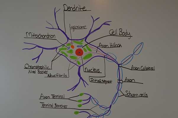
Structure of neurons is designed to optimize the flow of signals. Each neuron consists of a cell body, dendrites, and an axon. Dendrites receive incoming signals, while the axon carries information away from the cell body. This architecture ensures that communication is efficient and rapid, enabling the body to respond almost instantly.
Neurotransmitters play a critical role in this communication process. These chemical messengers are released at synapses, allowing signals to be passed from one neuron to another. The precise regulation of neurotransmitters ensures that messages are accurately transmitted, preventing disorders that can arise from imbalances.
Role of the Central Nervous System
The central part of the body’s communication network plays a pivotal role in processing information and coordinating responses. This component acts as the control center, interpreting sensory input and sending out commands to regulate bodily functions. Without it, the body would be unable to respond to changes in the environment, maintain balance, or carry out voluntary and involuntary actions.
Core Functions of the Brain and Spinal Cord
The brain and spinal cord form the core of this network, with each part contributing specialized functions. The brain processes complex information, while the spinal cord acts as a major pathway for transmitting signals between the brain and other parts of the body. Together, these two organs enable the body to react swiftly and effectively to internal and external stimuli.
Key Responsibilities of the Central Control Center
The main responsibilities of the brain and spinal cord include regulating motor skills, managing sensory input, and maintaining vital functions like breathing and heartbeat. The brain also processes emotions, thoughts, and memory, integrating these elements to influence behavior and decision-making.
| Component | Function |
|---|---|
| Brain | Processes sensory data, controls emotions, thoughts, and memory |
| Spinal Cord | Transmits signals between the brain and peripheral organs, controls reflex actions |
| Cerebellum | Coordinates voluntary movements and maintains posture and balance |
| Brainstem | Regulates basic life functions such as heartbeat, breathing, and sleep cycles |
Peripheral Nervous System Overview
The peripheral network serves as a crucial link between the central control center and the rest of the body. It extends throughout the body, connecting limbs, organs, and tissues to the brain and spinal cord. This extensive network ensures that information flows efficiently in both directions, allowing for coordinated movements and responses to environmental changes.
Key Components of the Peripheral Network
This network consists of sensory and motor pathways that carry signals to and from the central control center. The sensory pathways collect information from various parts of the body and send it to the brain, while the motor pathways transmit instructions from the brain to muscles and glands. Below are the main components:
- Somatic Nerves: Responsible for voluntary control over skeletal muscles.
- Autonomic Nerves: Regulate involuntary functions such as heart rate, digestion, and breathing.
- Cranial Nerves: Emerging directly from the brain, they control functions related to sight, smell, taste, and facial muscles.
- Spinal Nerves: Transmit messages between the spinal cord and the body’s extremities, playing a key role in reflex actions.
Functions and Roles of Peripheral Pathways
Peripheral pathways allow for a wide range of bodily functions, from voluntary movements to automatic regulation of vital processes. These pathways ensure that the body can detect changes in the environment and respond accordingly. Below are some primary roles of the peripheral network:
- Sensory Input: Gathering data about the external environment and internal conditions.
- Motor Output: Sending signals from the brain to initiate muscle movement or glandular secretion.
- Autonomic Regulation: Maintaining balance in internal processes such as digestion, heart rate, and respiratory rhythm.
Exploring the Brain’s Structure and Functions
The brain is the central hub of control and coordination in the body, responsible for interpreting sensory information, regulating behaviors, and managing complex cognitive processes. Its structure is highly specialized, allowing it to perform a vast array of tasks essential for survival. Understanding the various regions of the brain and their functions is crucial to grasping how the body responds to internal and external stimuli.
Key Regions of the Brain
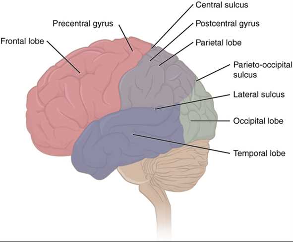
The brain is divided into several distinct areas, each responsible for different aspects of function. These regions work together to ensure smooth communication and efficient regulation of bodily processes. The main regions include:
- Cerebrum: The largest part, responsible for higher functions such as thought, memory, and voluntary movement.
- Cerebellum: Coordinates balance, posture, and fine motor skills, ensuring smooth and controlled movements.
- Brainstem: Controls basic life functions such as heartbeat, breathing, and sleep cycles.
Functions of the Brain’s Key Areas
Each part of the brain plays a critical role in maintaining both voluntary and involuntary functions. The cerebrum is involved in conscious actions, decision-making, and emotional responses, while the cerebellum ensures physical coordination. The brainstem, on the other hand, is vital for survival, overseeing essential functions like heart rate and respiration.
Neuroplasticity is another fascinating aspect of the brain. This term refers to the brain’s ability to adapt and reorganize itself by forming new neural connections in response to learning, experience, or injury. This remarkable property allows the brain to remain flexible and responsive throughout life.
How Nerve Impulses Travel Through the Body
The body relies on a highly efficient communication network that allows for the rapid transmission of electrical signals. These impulses carry vital information to various parts of the body, enabling it to respond to changes and maintain proper function. The process of how these messages travel is complex but essential for bodily coordination and reaction.
The Pathway of Nerve Impulses
When a signal is initiated, it follows a specific pathway, starting at a receptor or sensory organ and traveling through specialized cells before reaching the brain or spinal cord. The journey of a nerve impulse can be broken down into several key stages:
- Signal Reception: A stimulus, such as heat or touch, is detected by sensory receptors located in the skin or other organs.
- Signal Transmission: The receptor sends the signal to the nearest neuron, which acts as a messenger by transmitting the electrical impulse along its axon.
- Synapse Crossing: Once the signal reaches the end of the axon, it crosses the synapse (the gap between two neurons) by releasing neurotransmitters.
- Signal Relay: The neurotransmitter binds to the receptors of the next neuron, triggering the continuation of the impulse.
- Processing and Response: The signal eventually reaches the brain or spinal cord, where it is processed and a response is initiated, such as muscle movement or hormone secretion.
The Role of Myelin Sheath in Signal Speed
The myelin sheath, a fatty layer that surrounds certain nerve fibers, plays a crucial role in increasing the speed of signal transmission. It allows the electrical impulses to travel much faster by insulating the axon, reducing energy loss and preventing interference. This ensures that the body can respond to stimuli almost instantly.
- Faster Transmission: Myelinated fibers conduct impulses much faster than non-myelinated fibers, enhancing reflexes and quick responses.
- Efficiency: The insulation prevents signal degradation, ensuring clear communication between the brain, spinal cord, and peripheral organs.
Test Questions on Spinal Cord Function
The spinal cord is a crucial pathway for transmitting signals between the brain and the body, responsible for a variety of motor and sensory functions. It acts as a communication highway, enabling reflex actions and coordination. To understand its role, it’s important to evaluate the key functions and structures that support its vital processes.
Key Functions of the Spinal Cord
Before diving into specific questions, let’s review some of the core functions of the spinal cord:
- Signal Transmission: The spinal cord transmits information from the brain to the rest of the body and vice versa.
- Reflex Control: It plays a central role in reflex actions, allowing for quick, involuntary responses to stimuli.
- Motor Coordination: It helps coordinate voluntary movements, particularly for the limbs and trunk.
- Sensory Processing: The spinal cord carries sensory information, such as pain, temperature, and touch, to the brain for processing.
Common Questions to Assess Spinal Cord Function
Understanding how the spinal cord works involves asking questions that cover its anatomy and physiological roles. Below are some sample questions that can help evaluate comprehension:
- What is the primary function of the spinal cord?
The spinal cord’s primary function is to transmit signals between the brain and the rest of the body, as well as control reflexes. - How does the spinal cord contribute to reflex actions?
The spinal cord processes reflexes by allowing immediate responses to stimuli without involving the brain. - What is the role of the spinal cord in motor control?
The spinal cord assists in coordinating voluntary muscle movements, particularly for limbs and torso. - How does the spinal cord process sensory input?
Sensory information is relayed through sensory neurons to the spinal cord, which then sends it to the brain for interpretation. - What is the effect of spinal cord injury on body function?
Injury to the spinal cord can result in loss of motor skills, sensation, and reflex function below the site of damage.
These questions help assess knowledge on the spinal cord’s structure and its essential functions in the body. Understanding these elements is key to grasping how the body maintains coordination and responds to changes in the environment.
Autonomic Nervous System Explained
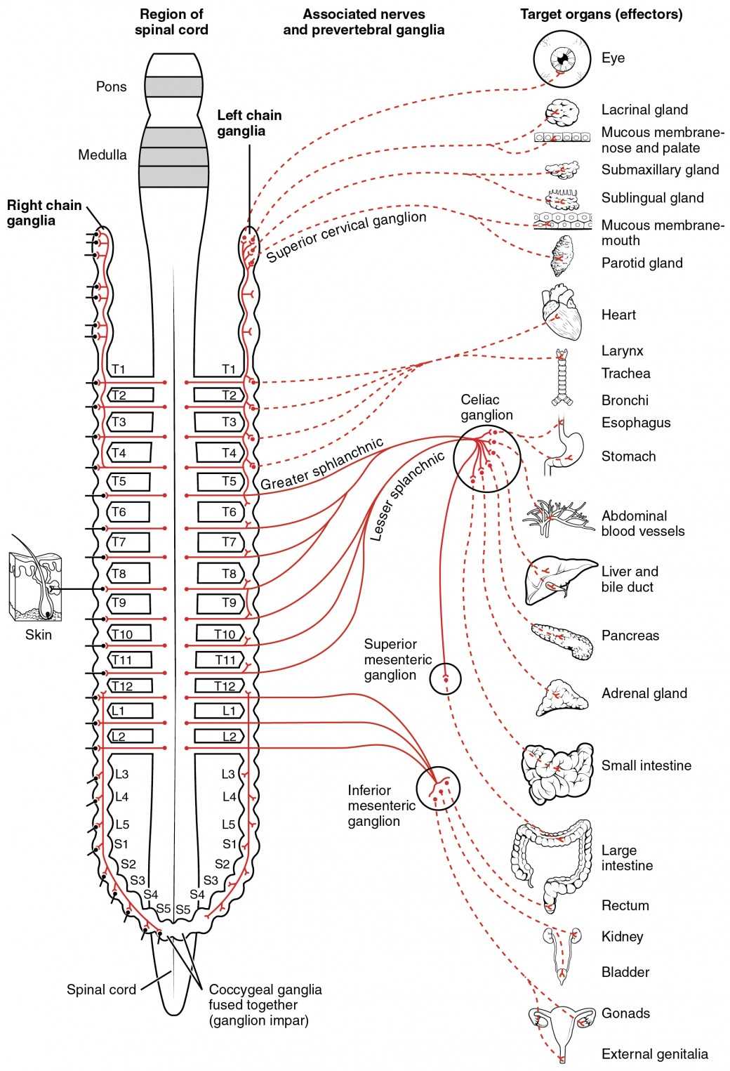
The body has a built-in mechanism that controls vital functions without conscious effort. This mechanism regulates processes such as heart rate, digestion, and respiratory rate, ensuring the body functions smoothly even when not actively thinking about it. Understanding this system is essential to grasp how the body maintains balance and responds to internal and external changes.
This particular network operates automatically, overseeing essential functions that are not under voluntary control. The body’s ability to perform these tasks efficiently and consistently is crucial for survival and homeostasis.
Key Functions of the Autonomic Network
The autonomic network is divided into two major branches, each responsible for different physiological processes:
| Branch | Primary Function |
|---|---|
| Sympathetic Branch | Prepares the body for “fight or flight” responses, increasing heart rate, dilating pupils, and redirecting blood flow to muscles. |
| Parasympathetic Branch | Promotes relaxation and recovery, slowing heart rate, stimulating digestion, and aiding in energy conservation. |
How It Regulates Bodily Functions
This network functions by transmitting signals through specialized pathways to organs and tissues. The brain and spinal cord coordinate with various organs to manage stress, rest, digestion, and more. For instance, when stressed, the sympathetic branch activates a series of responses that prepare the body for action. Conversely, during rest, the parasympathetic branch helps the body calm down, promoting digestion and energy storage.
Understanding how the autonomic network works allows for deeper insights into how the body maintains equilibrium, ensuring it reacts appropriately to various stimuli, both internal and external, without conscious effort.
Motor vs. Sensory Pathways in Detail
The body relies on complex networks to relay information between the brain and various parts of the body. Two primary pathways are responsible for this communication: one controls voluntary actions, while the other manages sensory feedback from the environment. Understanding the distinction between these two pathways is essential for comprehending how the body coordinates movement and interprets external stimuli.
Motor pathways direct signals from the brain to muscles, enabling movement. Sensory pathways, on the other hand, transmit sensory information from the body back to the brain, allowing for the perception of touch, temperature, pain, and other sensory inputs. These pathways work in harmony to facilitate appropriate responses to stimuli and to ensure coordinated movement and awareness.
Motor Pathways: Controlling Voluntary Movements

Motor pathways begin in the brain and extend through the spinal cord to muscles throughout the body. These pathways allow for voluntary actions, such as walking, talking, and picking up objects. The motor pathway is divided into upper and lower motor neurons, each playing a specific role in movement control:
- Upper Motor Neurons: These neurons originate in the brain and transmit signals to the spinal cord, initiating movement.
- Lower Motor Neurons: These neurons carry signals from the spinal cord to the muscles, triggering muscle contraction and movement.
Damage to either part of the motor pathway can result in motor deficits, such as paralysis or weakness, depending on the location of the injury.
Sensory Pathways: Relaying Sensory Information
Sensory pathways are responsible for carrying information from sensory receptors to the brain, enabling the body to perceive touch, temperature, pain, and pressure. These pathways involve a series of neurons that transmit signals from the sensory organs to the brain for interpretation:
- First-order Neurons: These neurons receive sensory input from receptors in the skin, muscles, or organs and transmit it to the spinal cord.
- Second-order Neurons: These neurons relay the information from the spinal cord to the brainstem or thalamus, where the signal is further processed.
- Third-order Neurons: These neurons carry the signal from the thalamus to specific areas of the brain, allowing for conscious perception of the sensation.
Impairment of sensory pathways can lead to sensory deficits such as numbness or inability to perceive certain stimuli, depending on the area affected.
Both motor and sensory pathways are integral to the proper functioning of the body. They work together to enable movement and perception, providing a seamless interaction between the brain and the rest of the body. Understanding their structure and function helps clarify how the body coordinates responses to the environment and ensures effective motor control and sensory awareness.
Important Reflex Arcs to Remember
Reflex arcs are essential for the body’s ability to respond quickly to stimuli without the need for conscious thought. These rapid, automatic responses help protect the body from harm and maintain homeostasis by enabling immediate reactions to changes in the environment. Reflexes are controlled by neural pathways that involve sensory input, processing in the spinal cord, and motor output, all working in tandem to produce a fast response.
While many reflex arcs are simple and serve protective functions, others are more complex and contribute to various bodily functions. Recognizing the different types of reflex arcs can help in understanding how the body reacts to both external and internal stimuli without delay.
Here are some important reflex arcs to remember:
- Patellar Reflex (Knee-Jerk Reflex): This is a classic example of a monosynaptic reflex. It occurs when the patellar tendon is tapped, causing a brief stretch in the quadriceps muscle. The stretch activates sensory neurons, which then send a signal to the spinal cord, triggering motor neurons that cause the quadriceps to contract, resulting in the knee jerking forward.
- Withdrawal Reflex (Flexor Reflex): This reflex helps protect the body from injury. When a painful stimulus, such as touching something hot, is detected by sensory receptors in the skin, sensory neurons transmit the signal to the spinal cord. In response, the spinal cord sends signals through motor neurons to pull the affected body part away from the source of pain.
- Crossed Extensor Reflex: This reflex works in conjunction with the withdrawal reflex. When one leg withdraws due to a painful stimulus, the opposite leg extends to maintain balance and support the body’s weight. This helps prevent a fall when the body reacts to pain.
- Babinski Reflex: The Babinski reflex is typically observed in infants or in adults with certain neurological conditions. When the sole of the foot is stroked, the big toe extends upward and the other toes fan out. In adults, this reflex is abnormal and indicates potential damage to the central nervous pathway.
- Golgi Tendon Reflex: This reflex helps protect muscles from excessive force. When muscle tension increases to a dangerous level, sensory receptors called Golgi tendon organs in the tendons send signals to the spinal cord, causing the muscle to relax to prevent injury.
These reflexes highlight the body’s ability to respond quickly and efficiently to stimuli, often without the need for conscious awareness. Understanding reflex arcs and their roles helps in recognizing how the body maintains safety and balance through automatic responses.
Neurotransmitters and Their Impact
Neurotransmitters are chemical messengers that play a crucial role in communication between neurons. They enable the transmission of signals across synapses, influencing various functions such as mood, movement, and cognition. These chemicals are released by neurons and bind to receptors on adjacent cells, triggering a wide range of responses within the body. Understanding the impact of neurotransmitters is key to understanding how the brain and other organs interact and maintain homeostasis.
Each neurotransmitter has a specific function and can have varying effects depending on the area of the body they act upon. Some neurotransmitters promote excitability in target cells, while others inhibit their activity. The balance between these chemicals is essential for proper functioning, and imbalances can lead to a variety of neurological and psychological disorders.
Common Neurotransmitters and Their Functions
| Neurotransmitter | Primary Function | Associated Disorders |
|---|---|---|
| Acetylcholine | Regulates muscle movement and memory formation. | Alzheimer’s disease, myasthenia gravis. |
| Dopamine | Influences mood, motivation, and motor control. | Parkinson’s disease, schizophrenia. |
| Serotonin | Affects mood, appetite, and sleep regulation. | Depression, anxiety, insomnia. |
| Norepinephrine | Involved in alertness, arousal, and stress responses. | Depression, ADHD, anxiety disorders. |
| GABA (Gamma-Aminobutyric Acid) | Inhibits neuron activity, reducing anxiety and promoting relaxation. | Seizures, anxiety disorders. |
Impact of Imbalances
Imbalances in neurotransmitter levels can have profound effects on health. For example, reduced dopamine levels are associated with Parkinson’s disease, which affects movement control. On the other hand, low serotonin levels are linked to mood disorders like depression. Similarly, excessive glutamate can lead to excitotoxicity, damaging neurons and contributing to conditions such as Alzheimer’s disease and stroke.
In addition to their role in disorders, neurotransmitters are also critical in everyday processes such as learning, memory, and emotional regulation. Understanding how these chemicals work allows for better insight into how the body functions at the cellular level and offers potential targets for therapeutic interventions.
The Blood-Brain Barrier and Its Importance
The blood-brain barrier (BBB) is a critical protective mechanism that separates the brain’s delicate tissues from harmful substances in the bloodstream. Its primary role is to maintain a stable environment for the brain by preventing toxic materials and pathogens from entering while allowing essential nutrients to pass through. This selective permeability ensures the brain functions optimally and remains shielded from potential damage.
Comprised of tightly joined endothelial cells, the blood-brain barrier acts as a filter, allowing only specific molecules to pass through. While this offers immense protection, it can also complicate the delivery of certain medications to the brain. Understanding the BBB’s function is crucial for developing treatments for neurological disorders and understanding the mechanisms behind certain diseases.
Key Functions of the Blood-Brain Barrier
- Protection: Blocks harmful substances like toxins, viruses, and bacteria from reaching the brain.
- Selective Transport: Allows only certain nutrients, such as glucose and amino acids, to enter the brain.
- Maintains Homeostasis: Regulates the environment of the brain by controlling ion balance and preventing fluctuations in neurotransmitter levels.
Factors Affecting the Blood-Brain Barrier
- Age: The barrier may become more permeable with age, increasing vulnerability to diseases.
- Infection: Infections can temporarily compromise the blood-brain barrier, allowing pathogens to enter the brain.
- Diseases: Conditions like Alzheimer’s disease, stroke, or multiple sclerosis can damage the barrier, leading to potential neuroinflammation or other complications.
The ability to bypass or enhance the function of the blood-brain barrier is a major focus in the field of medicine, particularly in developing treatments for neurological conditions. Research into this area aims to improve drug delivery methods to the brain, providing better solutions for disorders that currently have limited treatment options.
Diseases Affecting the Nervous System
Various disorders can impact the body’s communication network, leading to significant changes in functionality and quality of life. These conditions often involve disruptions in the pathways that control movement, sensation, cognition, and overall bodily function. Understanding how these illnesses affect the brain, spinal cord, and peripheral pathways is essential for diagnosis, treatment, and management strategies.
Some conditions result from genetic factors, while others arise due to infections, trauma, or environmental influences. The severity and progression of these disorders vary, but many can lead to long-term challenges for affected individuals. Early detection and intervention are crucial in slowing or managing their effects.
Common Disorders
- Alzheimer’s Disease: A degenerative disorder that impairs memory, thinking, and behavior, often linked to cognitive decline.
- Parkinson’s Disease: A progressive disorder that affects movement, causing tremors, rigidity, and slow motor skills.
- Multiple Sclerosis: An autoimmune condition where the immune system attacks the protective coverings of nerve fibers, leading to inflammation and damage.
- Stroke: Occurs when blood flow to the brain is interrupted, leading to brain cell damage and various neurological impairments.
- Epilepsy: A neurological disorder marked by recurring seizures due to abnormal brain activity.
Factors Contributing to Neurological Disorders
- Genetic Predisposition: Some conditions, such as Huntington’s disease, are inherited and can lead to progressive dysfunction.
- Trauma: Physical injuries, such as concussions or spinal cord damage, can cause lasting neurological effects.
- Infections: Viral or bacterial infections that affect the brain, like meningitis or encephalitis, can lead to severe complications.
- Environmental Factors: Exposure to toxins, chemicals, or radiation can increase the risk of developing certain disorders.
Research into the causes, effects, and treatments for these conditions is ongoing. Advances in medicine continue to provide new insights into how to better manage, prevent, or potentially reverse some of the damage caused by these diseases.
Practical Applications of Nervous System Knowledge
Understanding how the body’s communication network operates is not only essential for medical fields but also offers practical solutions in everyday life. The insights gained from studying this system can be applied across a wide range of disciplines, from healthcare and rehabilitation to technology and education. By exploring how the brain, spinal cord, and peripheral pathways function, experts are able to develop targeted interventions that improve quality of life, enhance performance, and promote overall well-being.
In healthcare, for instance, this knowledge is fundamental for diagnosing and treating a variety of conditions. It allows for more effective treatment of injuries, diseases, and disorders related to the body’s communication infrastructure. Likewise, advances in rehabilitation rely heavily on understanding how the brain and other organs interact to restore function.
Medical Applications
- Neurorehabilitation: Knowledge of how the brain recovers from injury or illness helps therapists develop more effective recovery programs for patients with stroke, spinal cord injury, or traumatic brain injuries.
- Neurosurgery: Surgeons use this knowledge to operate on conditions such as tumors, aneurysms, or congenital abnormalities affecting brain or spinal cord structures.
- Pain Management: Understanding pain pathways aids in developing treatments for chronic pain conditions, such as neuropathy, by targeting specific receptors or neural circuits involved in pain perception.
Technological Innovations
- Brain-Computer Interfaces: Advances in this area allow people with severe disabilities to control prosthetic limbs or communicate using only neural signals, bridging the gap between biology and technology.
- Neuroprosthetics: Devices that interact with the brain to assist in movement or sensory function are improving the lives of individuals with amputations or neurological disorders.
- Artificial Intelligence: Understanding cognitive processes in humans is aiding the development of more sophisticated AI, capable of mimicking aspects of human decision-making and problem-solving.
Beyond medical and technological fields, this knowledge also plays a critical role in enhancing cognitive performance, improving education methods, and ensuring workplace safety. From optimizing learning techniques to understanding stress responses, the practical benefits of understanding the body’s communication system are far-reaching.
Reviewing Test Strategies for Nervous System Topics
When preparing for assessments related to the body’s communication network, it is crucial to adopt effective strategies that ensure a deep understanding of key concepts. A structured approach to studying can help individuals retain information more efficiently and apply their knowledge effectively during evaluations. From mastering fundamental principles to applying complex processes, several techniques can enhance one’s readiness for challenges related to the body’s communication pathways.
First, it is essential to break down complex topics into smaller, manageable sections. Focusing on one concept at a time allows for better retention and understanding. Visual aids such as diagrams or models can help clarify how various components interact, providing a clear overview of their functions and relationships. Another useful strategy is active recall, where individuals test their memory by trying to explain a concept without referring to notes, reinforcing what they have learned.
Another effective strategy is to use practice scenarios or questions that mirror the style of the evaluation. This technique not only helps identify weak areas but also builds confidence in applying theoretical knowledge to practical situations. Collaboration with peers can also be beneficial, as explaining complex topics to others reinforces understanding while also offering different perspectives.
Finally, regular review sessions are key to long-term retention. Revisiting previously studied material periodically helps prevent forgetting and solidifies the connections between concepts. Whether through flashcards, summary notes, or group discussions, consistent reinforcement ensures that knowledge remains fresh and accessible when needed most.
Common Mistakes in Nervous System Tests
When assessing knowledge of the body’s communication network, there are several common pitfalls that can hinder performance. These errors often arise from misunderstandings or oversights during the preparation or assessment process. Recognizing and addressing these mistakes can greatly improve results and ensure a more thorough grasp of essential concepts.
One frequent mistake is misinterpreting terminology. Many individuals confuse related terms that describe different processes or components, which can lead to confusion in both multiple-choice and written response questions. Another common issue is neglecting the importance of understanding relationships between different parts of the body’s communication network, leading to incomplete answers or missing critical connections.
Other errors occur when students focus too heavily on memorization rather than comprehension. Rote learning without truly understanding the underlying mechanisms can result in the inability to apply knowledge in real-world scenarios. This can be especially problematic in questions that require the application of concepts to unfamiliar situations.
- Overlooking small details: Even seemingly minor details can affect the accuracy of an answer, such as forgetting the role of specific neurotransmitters or the function of particular pathways.
- Not understanding cause and effect: Failing to link cause-and-effect relationships between different components of the body’s network often results in incomplete or inaccurate explanations.
- Underestimating complexity: Many underestimate the complexity of certain processes, leading to oversimplified answers that miss key nuances.
- Not practicing with mock questions: Without using practice questions or scenarios, it’s difficult to identify which areas need improvement and which concepts require further study.
Avoiding these mistakes involves adopting strategies that encourage deeper learning, such as using active recall, applying knowledge to practical examples, and reviewing concepts regularly. A strong focus on understanding the “why” behind each process is crucial for success in assessments.