Onion Root Tip Mitosis Lab Worksheet Answers
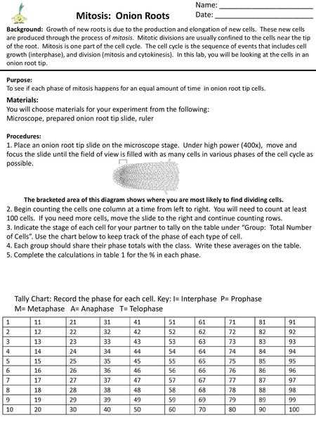
The study of cell division is fundamental to understanding how living organisms grow, develop, and reproduce. Observing how cells split and form new ones provides valuable insights into the intricate processes that sustain life. This topic focuses on a specific example from plant cells, allowing students to closely examine the stages of cellular replication under a microscope.
During this exercise, the key phases of cellular division are identified and analyzed through carefully prepared slides. By examining the changes occurring within plant cells, students can learn how the process of division ensures proper growth and the continuation of life. The following sections explore the methods used to study these cellular activities, along with the expected findings and interpretations of results.
Cell Division Process Overview
The process of cell division is a crucial event that occurs in living organisms, ensuring the formation of new cells for growth, repair, and reproduction. In plant cells, this division follows a series of well-defined stages, each playing a specific role in the creation of two genetically identical cells. By observing these stages, we can gain a deeper understanding of how organisms maintain and renew their cellular structures.
In this study, we focus on a specific part of the plant where cell division is most active. By examining these cells under a microscope, the distinct phases of division become observable. Each phase has unique characteristics that allow researchers to track and identify the progression of cell division.
| Stage | Key Characteristics |
|---|---|
| Prophase | Chromosomes condense, nuclear membrane begins to break down. |
| Metaphase | Chromosomes align at the cell’s center, spindle fibers form. |
| Anaphase | Chromatids are pulled toward opposite sides of the cell. |
| Telophase | Two new nuclear membranes form, chromosomes de-condense. |
| Cytokinesis | Cell membrane pinches to form two separate cells. |
Understanding these stages is essential for studying cell growth, development, and the overall functioning of living organisms. By observing plant cells, we can better grasp the complexity and precision involved in cellular division.
Understanding Cell Division Stages in Detail
Cell division is a highly organized process that ensures the accurate duplication of genetic material and the proper formation of new cells. The entire process is divided into distinct phases, each with specific events that contribute to the overall division. Understanding each stage in detail helps clarify how organisms grow and how tissues regenerate. Below is an exploration of the key phases involved in cellular replication.
Prophase and Metaphase
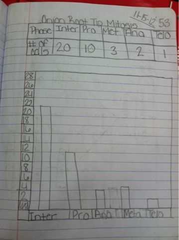
In the first stages of division, the cell prepares for the separation of its genetic material. During prophase, the chromosomes become visible as they condense, and the nuclear membrane begins to dissolve. Spindle fibers, which play a crucial role in separating chromosomes, start to form. In the subsequent stage, metaphase, the chromosomes align at the cell’s center, ensuring that they are evenly divided during the next phase. This alignment is essential for maintaining genetic integrity in the daughter cells.
Anaphase and Telophase
Once the chromosomes are aligned, the cell proceeds into anaphase, where the chromatids (individual strands of chromosomes) are pulled toward opposite poles of the cell. This separation ensures that each new cell will receive an identical set of genetic material. Following this, telophase occurs, where two new nuclear membranes form around the separated chromosomes, signaling the near end of the division process. The chromosomes begin to de-condense, returning to their less visible form.
Each of these stages plays a vital role in ensuring the accuracy and efficiency of cellular replication, making them fundamental to the overall health and growth of organisms.
How Plant Cells Demonstrate Cell Division
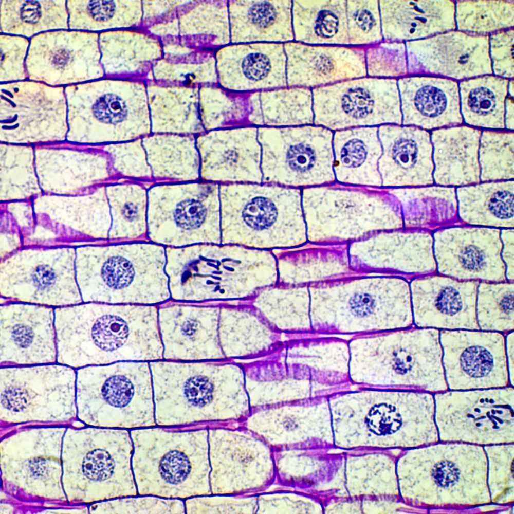
Certain plant tissues provide an excellent opportunity to observe the process of cell division due to their high rate of cellular activity. These tissues are often found at the tips of growing parts of the plant, where new cells are continually produced. By examining these areas under a microscope, it becomes possible to clearly see the different phases of cellular replication in action. The process is made more visible because the cells are actively dividing, making it easier to identify each stage of the division cycle.
In particular, the cells within the actively growing regions of the plant display clear signs of division. As cells progress through various stages, such as the condensation of chromosomes and the formation of new nuclei, their distinct characteristics can be observed under magnification. This visual demonstration not only allows students to identify these stages but also deepens the understanding of how cells maintain and grow through division.
By focusing on plant cells in these rapidly dividing areas, it’s possible to witness the steps of cellular reproduction in real time, offering a tangible connection to the broader biological processes that drive growth and development in plants.
Key Observations During Cell Division Process
Observing cell division involves noting several crucial changes that occur within the cell. These changes, which mark the different phases of division, can be identified by their distinct visual characteristics. By closely examining the cells under a microscope, it becomes clear how the genetic material is organized, separated, and distributed to form two identical cells. This process plays a fundamental role in the growth, repair, and reproduction of organisms.
Chromosome Behavior
One of the most noticeable aspects of cell division is the behavior of the chromosomes. Initially, they condense into tightly packed structures, becoming visible under a microscope. This condensation allows for the accurate separation of genetic material during later stages. As division progresses, the chromosomes align in the cell’s center and then move toward opposite poles, ensuring that each new cell will inherit an identical set of genetic information.
Formation of New Cells
As the cell division process nears completion, the final step involves the physical separation of the two daughter cells. This is observed as the cell membrane pinches and forms two distinct, fully separated cells. The development of new nuclei around the separated chromosomes marks the end of division and the beginning of the cell’s normal function within the organism.
These key observations offer a clear understanding of how the intricate process of cell division ensures the continuity of life through the formation of new, genetically identical cells.
Identifying Phases in Plant Cells
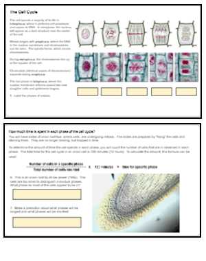
Cell division in plants follows a structured sequence of stages, each with distinct visual markers that help identify the specific phase the cell is undergoing. By carefully observing cells under a microscope, it’s possible to recognize these stages and understand how the division process ensures the growth and regeneration of plant tissues. Recognizing each phase allows for a deeper understanding of the biological mechanisms behind cellular reproduction.
When observing plant cells, the key phases to identify include the following:
- Prophase: Chromosomes begin to condense and become visible. The nuclear membrane starts to break down, and spindle fibers begin to form.
- Metaphase: Chromosomes align at the center of the cell, forming a plate. This arrangement is essential for the proper separation of genetic material.
- Anaphase: The chromatids are pulled apart and move toward opposite poles of the cell, ensuring that each new cell will receive a complete set of genetic material.
- Telophase: New nuclear membranes begin to form around the separated chromatids, and the chromosomes start to de-condense.
- Cytokinesis: The final step, where the cell membrane pinches off, creating two distinct daughter cells, each with a full set of genetic information.
By understanding and recognizing these stages, one can gain insight into the dynamic process of cellular division and its essential role in maintaining plant growth and health.
Using a Microscope to Study Cell Division
A microscope is an essential tool for examining the detailed processes of cell division, especially in plant cells. By magnifying the cells, students and researchers can observe the various stages of the division process with clarity. Using the right magnification and proper technique allows for accurate identification of different phases and a deeper understanding of how cells replicate and grow.
To effectively study cell division, it is important to follow these steps when using a microscope:
- Prepare the Slide: Start by preparing a thin, well-stained slide to ensure clear visibility of the cell structures. Use appropriate dyes to highlight the chromosomes and other cellular components.
- Adjust the Magnification: Begin with a lower magnification to locate the cells, then gradually increase it to clearly see the details of the cell’s internal structures.
- Focus Properly: Carefully adjust the focus to bring the cells into sharp view. Fine focusing is essential for observing the different stages of division with precision.
- Identify Key Features: Look for key indicators of the different stages of division, such as the condensation of chromosomes in the early phases and the separation of chromatids during later stages.
- Record Observations: Take note of the time spent observing each phase and record any significant observations about the behavior of the cells. This will help in comparing and analyzing the results later.
Using a microscope not only allows for the observation of cellular processes but also enhances the understanding of how organisms grow and reproduce at the cellular level.
Why Certain Plant Tips Are Ideal for Study
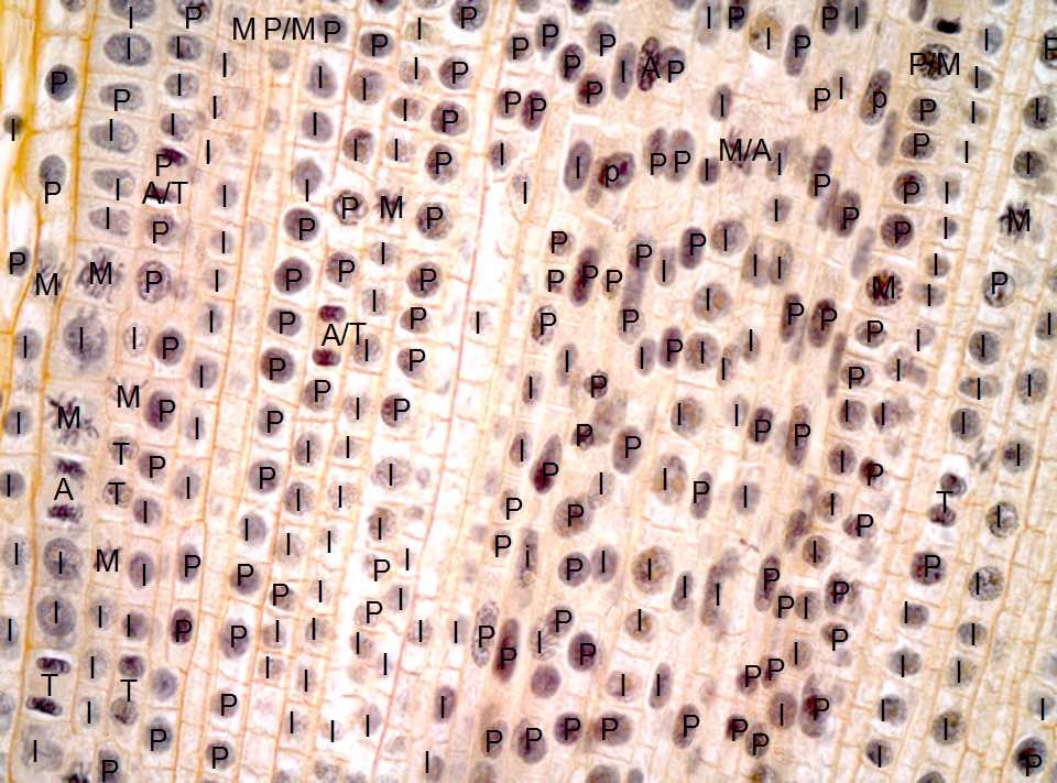
Certain plant tissues, especially those found at the growing ends of roots, offer a unique opportunity to study cell division due to their rapid cellular activity. These regions contain cells that are actively undergoing division, making them an ideal subject for examining the stages of cellular reproduction. The high frequency of dividing cells in these areas allows researchers and students to observe all phases of cell division in real-time.
Rapid Cell Division
One of the key reasons these particular plant tips are ideal for study is the speed at which the cells divide. The growing tips of plants are characterized by a high rate of mitotic activity, meaning that many cells are in the process of division at any given time. This allows for a higher likelihood of observing cells in various stages of division under the microscope, providing a comprehensive view of the process.
Clear Visibility of Dividing Cells
Additionally, the structure of the tissue makes it easier to observe cellular changes. The cells are generally larger, and the division stages are more easily distinguishable. Because the cells are tightly packed in the growth zone, they provide a clear view of the changes in chromosome arrangement, the formation of spindle fibers, and other key features of cellular division.
These factors combined make plant tips perfect for studying cellular processes like growth, repair, and reproduction, offering a clear and accessible means of observing fundamental biological mechanisms.
Preparing Slides for Cell Division Observation
Creating a well-prepared slide is crucial for effectively observing the stages of cell division under a microscope. The process involves selecting a suitable sample, preparing it correctly, and ensuring it is stained for optimal visibility. Proper slide preparation allows for the clear identification of the various phases of cell division, enabling accurate study and analysis.
Collecting the Sample
The first step in preparing a slide is to collect a sample from the plant tissue. Choose a region where cells are actively dividing, such as the tips of growing plant parts. These areas provide a high concentration of dividing cells, allowing for a better chance of observing all stages of the division process. Once the sample is collected, it must be carefully handled to preserve the cells for microscopic examination.
Staining and Mounting the Sample
After the sample is prepared, it is important to stain the cells to enhance the contrast and visibility of internal structures such as chromosomes. Staining agents, such as iodine or acetocarmine, are commonly used to make the genetic material stand out. Once stained, the sample is carefully mounted on a glass slide with a coverslip, ensuring that the cells are flat and evenly spread out for clear observation. Avoiding air bubbles during this process is essential to prevent obscuring the view under the microscope.
By following these steps, you can create a well-prepared slide that allows for the detailed observation of cellular processes, offering insights into the mechanisms of growth and reproduction in plants.
Common Mistakes in Cell Division Studies
During the study of cellular processes, particularly the stages of cell division, there are several common errors that can affect the quality of observations and results. These mistakes often arise from improper technique, incorrect preparation, or a lack of attention to detail. Understanding and avoiding these common pitfalls is crucial for ensuring accurate and reliable outcomes when observing cell division under a microscope.
Improper Slide Preparation
One of the most frequent mistakes in preparing slides for observation is not properly preparing the sample. If the sample is too thick, it can obscure the view, making it difficult to identify individual cells. Additionally, if the staining process is done incorrectly, the cells may not be clearly visible, making it hard to differentiate between the different phases of division. Properly preparing the slide, ensuring a thin and even sample, and using the correct amount of stain are all essential steps for clear observation.
Incorrect Microscope Settings
Another common issue is not adjusting the microscope properly. If the magnification is too low or too high, it can either fail to show the necessary details or make it difficult to focus on the cells. The focus should be adjusted gradually, and fine-tuning is often required to achieve the clearest image. Additionally, improper lighting can also affect visibility, so it’s important to ensure that the light is at the correct intensity for the sample being observed.
By avoiding these mistakes, the study of cell division becomes more accurate, allowing for a better understanding of the process and its significance in growth and reproduction.
| Common Mistake | Solution |
|---|---|
| Thick sample preparation | Ensure the sample is thin and even to allow clear viewing of cells. |
| Improper staining | Use the correct amount and type of stain to enhance cell visibility. |
| Incorrect microscope settings | Adjust magnification and focus carefully to obtain sharp images. |
| Insufficient lighting | Ensure proper lighting intensity for the clarity of the sample. |
How to Record Cell Division Findings Accurately
Accurately documenting observations during the study of cellular processes is essential for drawing reliable conclusions and ensuring reproducibility. Properly recording findings involves more than just noting what is seen; it requires attention to detail and systematic organization. Clear, precise documentation helps in comparing results and making valid interpretations of the stages observed in the cell division process.
To begin, it’s important to maintain consistency in how observations are recorded. Use a standardized format for noting the stages of cell division, such as identifying and categorizing cells at each phase. Record the number of cells observed at each stage, as well as any abnormalities or variations that might be present. It’s also crucial to describe the appearance of the cells, such as the arrangement of chromosomes or the presence of spindle fibers, as these features can indicate specific phases of division.
Another key aspect of accurate recording is timing. Since cell division occurs over time, it’s important to note the duration of each phase and the overall progression of the process. This can be especially useful when analyzing how long each phase takes and if there are any delays or irregularities in the division process.
Lastly, always cross-reference your observations with established diagrams or reference materials to ensure accuracy. This will help confirm that the findings align with known scientific principles and definitions. By following these practices, you can ensure that your recorded findings are reliable and valuable for further analysis.
Analyzing Cell Division Data from the Study
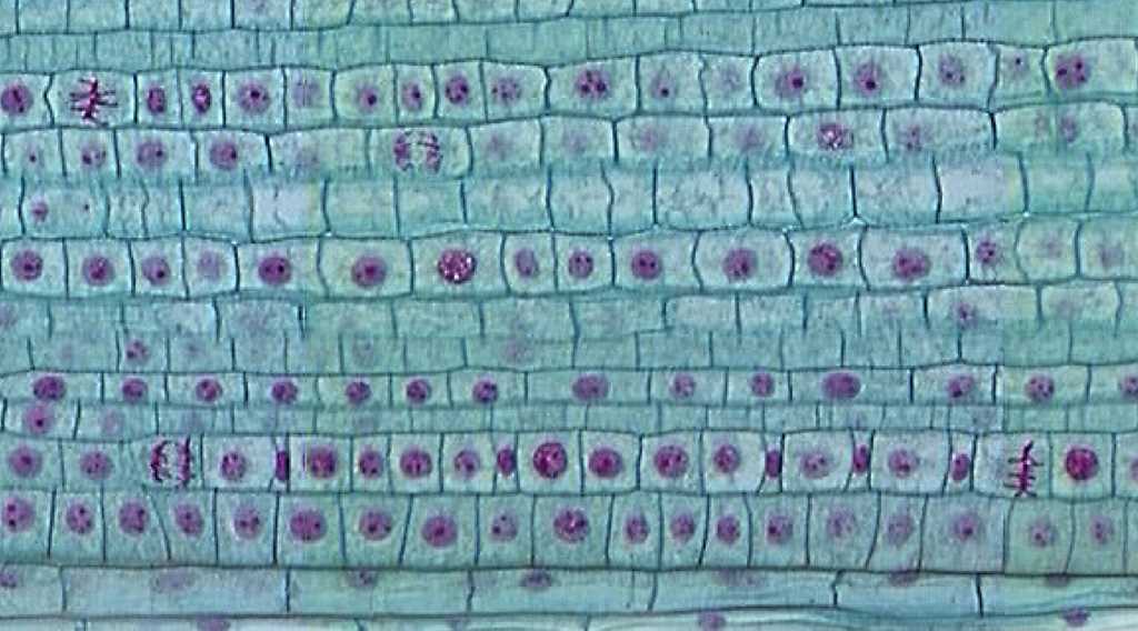
Once the data has been collected from the cellular division observations, the next step is to carefully analyze the findings. Proper analysis allows you to identify trends, determine the duration of each division phase, and assess the accuracy of the observed results. By reviewing the data methodically, you can draw meaningful conclusions about the efficiency and consistency of cell division processes in the sample studied.
Steps for Analyzing the Data
- Organize the Observations: Begin by sorting the recorded data into categories based on the observed stages. This helps in understanding the distribution of cells across different phases and identifying any irregularities.
- Calculate Cell Distribution: For each stage of cell division, calculate the percentage of cells observed. This will give you an insight into how many cells are in each phase at a given time and whether the division process is proceeding at a normal rate.
- Compare with Expected Results: Cross-reference your data with expected patterns of cell division, ensuring that the duration and sequence of phases are in line with known biological standards.
Interpreting Results
- Identify Abnormalities: Look for any unexpected patterns or irregularities, such as an imbalance in the number of cells in specific phases. These could indicate issues like cell cycle disruption or external factors affecting the process.
- Determine Phase Duration: Assess the relative lengths of each stage by analyzing how long cells remain in each phase. Variations in timing can provide insights into the overall health and efficiency of the division process.
- Draw Conclusions: After thorough analysis, summarize your findings. Are there any trends or anomalies that warrant further investigation? This step is crucial for understanding how cells function and replicate under different conditions.
By following a structured approach to data analysis, you can gain a deeper understanding of the cellular processes and generate reliable results that contribute to broader scientific knowledge.
Cell Cycle vs Division Process in Plant Cells
Understanding the differences between the overall cell cycle and the specific process of cell division is essential when studying how cells reproduce. While both terms are often used interchangeably, they refer to distinct phases that contribute to the growth and replication of cells. The cell cycle includes a series of events that prepare a cell for division, while division itself is just one phase of the entire cycle.
The cell cycle consists of multiple stages, including interphase, where the cell grows, replicates its DNA, and prepares for division. Interphase itself is divided into three sub-phases: G1 (growth phase), S (synthesis phase, where DNA replication occurs), and G2 (final preparations for division). These phases ensure the cell has everything it needs before division begins. During division, the cell undergoes several steps to separate its duplicated genetic material into two identical daughter cells.
In contrast, the process of cell division, which is often observed and analyzed in studies, typically refers to the stages of actual separation–such as prophase, metaphase, anaphase, and telophase. This process ensures that each daughter cell receives an exact copy of the original cell’s genetic material. While the cell cycle encompasses both the preparatory stages and the division stages, the latter is where visible changes in cell structure and chromosome movement occur.
In summary, while the cell cycle is a broader concept, including growth, DNA replication, and division, the division process itself is just a part of this continuous cycle. Understanding the distinction between these two is crucial for interpreting how cells grow and replicate in any organism, including plants.
Common Questions About Cell Division Observations
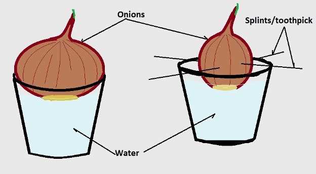
When studying cell division, especially in controlled settings, several questions often arise. These inquiries can range from the basic steps of the process to the specifics of how to effectively observe and interpret data. Understanding these common questions can enhance the learning experience and lead to more accurate observations.
Frequently Asked Questions
| Question | Answer |
|---|---|
| What is the best way to observe cell division? | Using a microscope to examine prepared slides is the most common method. It’s important to adjust the magnification to clearly see the stages of cell division. |
| How can I distinguish between different phases? | Look for specific changes in cell structure: chromosomes condense in prophase, align in metaphase, separate in anaphase, and begin to reform in telophase. |
| Why do cells go through division? | Cells divide to reproduce and generate new cells for growth, repair, and asexual reproduction. It ensures that the genetic material is evenly distributed between two new cells. |
| Can I speed up the process of division? | No, the timing of cell division is determined by biological factors. However, providing optimal environmental conditions like temperature and nutrient availability may influence the rate. |
| What is the significance of studying cell division? | Studying this process helps us understand how organisms grow, how tissues repair themselves, and how genetic material is passed on to the next generation. |
Addressing these questions can clarify the process and improve the accuracy of your observations. By becoming familiar with the key concepts, you will be better prepared to analyze and interpret the data collected from cell division studies.
How to Distinguish Between Cell Division Phases
Understanding the stages of cell division is essential for observing how a single cell splits into two. Each phase has distinct features that help identify the progression of division. By carefully examining the changes in the cell’s structure, it becomes easier to differentiate between the various stages.
Key Features of Each Phase
Cell division is divided into several key phases, each marked by specific changes. Here are the most important indicators:
- Prophase: The chromosomes begin to condense and become visible under the microscope. The nuclear membrane starts to break down, and the spindle fibers begin to form.
- Metaphase: Chromosomes align in the middle of the cell, also known as the metaphase plate. This is the stage where chromosomes are most easily seen and counted.
- Anaphase: The sister chromatids are pulled apart toward opposite poles of the cell. The cell elongates during this stage as the chromatids separate.
- Telophase: The nuclear membranes start to reform around each set of chromosomes, and the cell begins to split into two. Chromosomes begin to de-condense, and the spindle fibers disappear.
- Cytokinesis: The final separation of the cytoplasm occurs, resulting in two distinct daughter cells, each with a full set of chromosomes.
Observational Tips
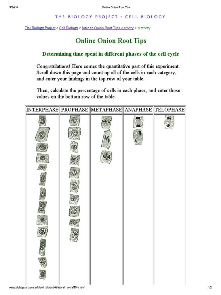
To effectively distinguish between these phases, focus on the following:
- Look for changes in chromosome shape and number as the cell progresses through each stage.
- Use the cell’s alignment and shape to determine if it is in metaphase or anaphase.
- Focus on the presence or absence of the nuclear membrane to identify telophase.
By closely examining the structural details and the sequence of changes, you will be able to identify and differentiate between the various phases of cell division with greater accuracy.
Practical Tips for Mitosis Lab Success
Performing experiments on cell division can be challenging, but with the right approach and careful attention to detail, you can achieve accurate results. Success in these experiments relies not only on understanding the theory but also on executing the practical steps effectively. Here are some essential tips to ensure a smooth and successful experiment.
Prepare Your Equipment Thoroughly
Before you begin the experiment, make sure all your tools are ready for use. This includes ensuring that your microscope is properly calibrated and that your slides and cover slips are clean. A well-prepared workspace can help avoid interruptions and mistakes during the observation process.
- Check the lens quality and focus of the microscope.
- Ensure slides are free from dust and air bubbles.
- Keep tissues or specimens moist to prevent them from drying out.
Stay Organized Throughout the Process
Keeping your work organized is key to achieving success. Label your slides clearly and maintain a systematic approach when documenting observations. This will make it easier to identify key stages and avoid confusion later on.
- Label each slide with a specific identifier and the date.
- Record observations immediately to ensure accurate data collection.
- Use a notebook or digital device to keep track of findings.
Focus on Key Cell Features
When analyzing the specimen, concentrate on the most important cellular changes. Understanding the key visual features of each phase will help you distinguish between them more easily. Pay close attention to the following:
- Chromosome alignment in the center of the cell.
- Changes in the nuclear membrane structure.
- Cell shape and separation during division.
Practice Patience and Precision
Observing the different stages of division requires patience. It may take time for the cells to progress through the stages, so be prepared to spend sufficient time under the microscope. Additionally, take precise measurements and notes to ensure your results are reliable.
- Take breaks if necessary to avoid fatigue during extended observation periods.
- Be methodical when documenting each stage and process observed.
By following these practical tips, you can enhance your chances of success in experiments involving cell division, ensuring that your results are clear, accurate, and meaningful.